Evelyn Pence
Medical Illustrator
ABOUT
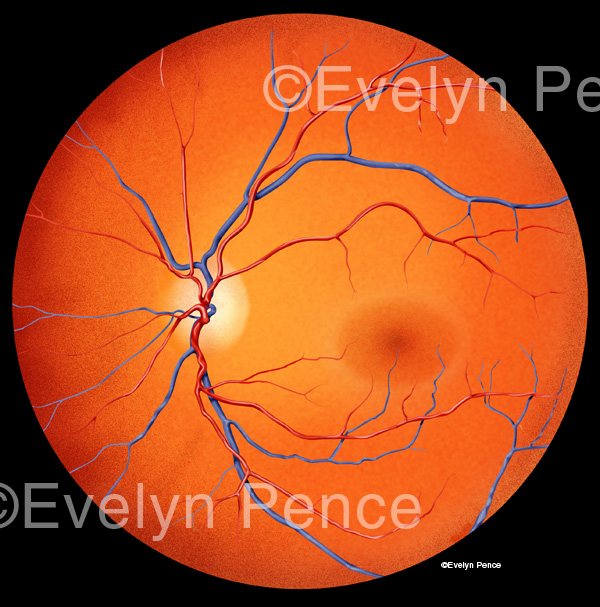
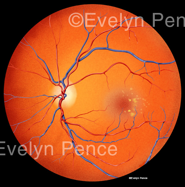
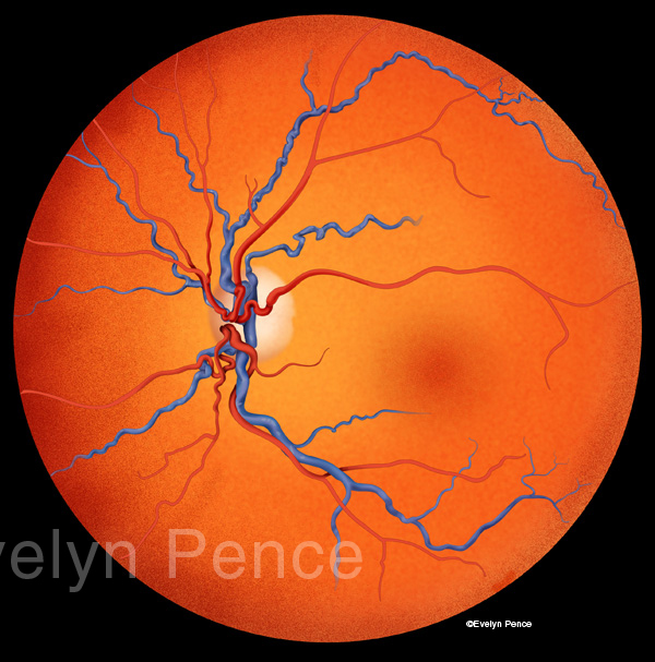
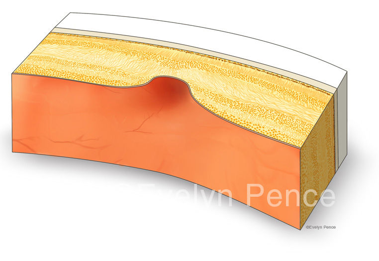
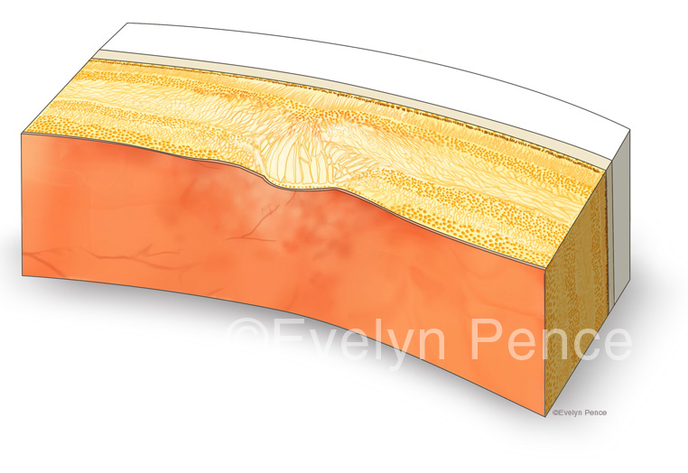
The human fundus (interior of the eye) was illustrated to compare healthy anatomy with two disorders associated with diabetic retinopthy: macula edema and central retinal vein occlusion, or CRVO. Because the presence of macular swelling is difficult to convey from the front view (as seen in the first 3 illustrations), two additional block diagrams showing a small cross-section of the fovea, one normal, one with macular edema, were created.